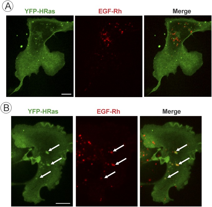
Fig. S6.
Analysis of colocalization of HRas and EGF-Rh in COS1 cells. COS1 cells transiently expressing YFP-HRas were incubated with 4 ng/mL EGF-Rh for 15 min at 37 °C. Single x–y confocal sections from the image z stacks acquired from cells expressing low (1.6× mV-HRas expression; A) or high levels (38× mV-HRas; B) of YFP-HRas are presented. Arrows indicate examples of colocalization of EGF-Rh and YFP-HRas in endosomes. (Scale bars: 10 μm.)

