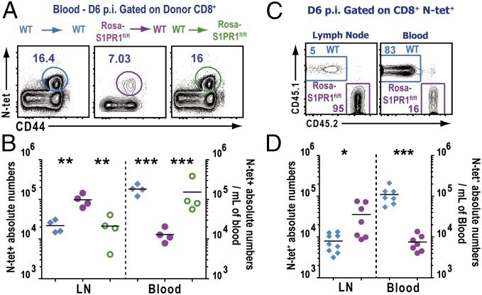
Fig. 3.
S1PR1 expression in hematopoietic cells is required for regulating effector T cell egress from the dLN after infection. Indicated BM chimeric mice were infected with VSV in the footpad, and the mice were administered tamoxifen orally starting at day 3 p.i. (A) Frequency of N-tet+ effector T cells in the blood of the indicated BM chimeric group at day 6 p.i. Dot plots represent congenic donor CD45+, CD8α+ gated cells. (B) Absolute numbers of N-tet+ effector T cells in the dLN and per milliliter of blood from the indicated chimeric groups WT→WT (blue), Rosa-S1PR1fl/fl→WT (purple), and WT→Rosa- S1PR1fl/fl (green). (C) Mixed BM chimeric mice were treated with tamoxifen, and, 6 d after VSV footpad infection, mice were killed. The distribution of the WT (blue gate) and S1PR1fl/fl (purple gate) N-tet+ T cell populations was assessed by FACS in the dLN and in blood. Numbers are frequencies of N-tet+ CD8 T cells present in each gate. (D) Absolute numbers of N-tet+ WT and S1PR1fl/fl in the dLN and in the blood (cells per mL) of tamoxifen-treated mixed BM chimeras at day 6 p.i. Data are representative of six independent experiments with at least four mice per group of chimeras. Horizontal lines indicate the mean. ***P < 0.0001 ; **P < 0.001; *P < 0.01.

