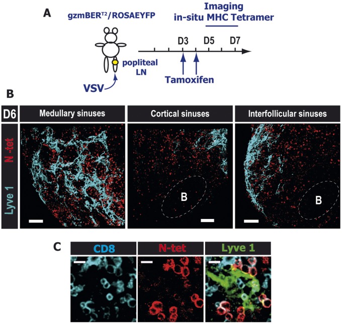
Fig. S5.
After a localized viral infection, endogenous antigen-specific CD8 T cells localize to the periphery of the dLN in the medullary and cortical/interfollicular lymphatic sinuses as they exit the LN. (A) Experimental scheme: Mice were infected in the footpad and subsequently treated twice orally with 1 mg of tamoxifen starting at day 3 p.i. (B) dLN were harvested, and thick sections (400 μm) were stained for N-Kb-tetramer in situ at 6 d p.i. Confocal images of N-specific T cells (red) in close proximity to medullary (Left), cortical (Center), and interfollicular (Right) sinuses. (Scale bars: 100 μm.) (C) Close-up of a cortical area showing N-tet+ effector CD8 T cells (red) in direct contact with Lyve1 (green) expressing lymphatics (Movie S1). (Scale bars: 10 μm.) B-cell follicles and T-cell zones were delineated using the B220 and CD8α staining, respectively (not shown). The data shown are representative of three different experiments with at least three mice per time point.

