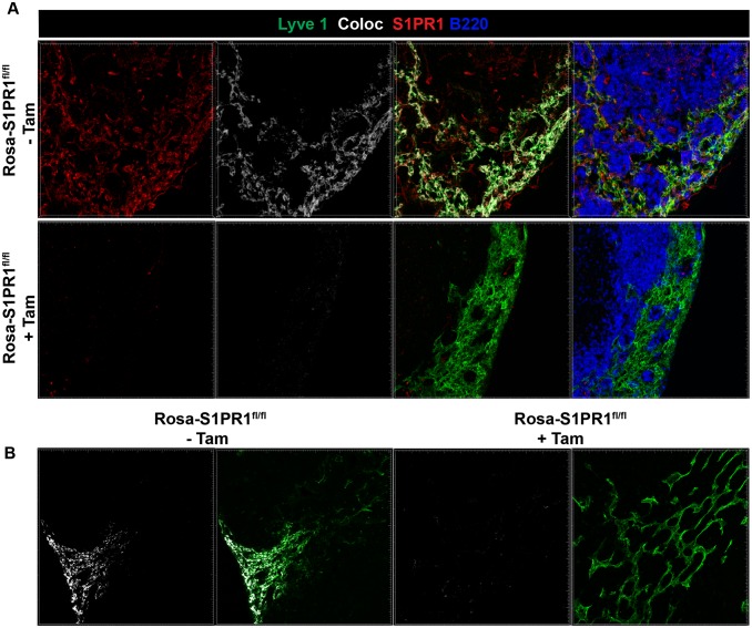
Fig. S6.
Efficiency of S1PR1 deletion in lymph nodes. Rosa-S1PR1fl/fl mice were either left untreated (−Tam) or treated with tamoxifen (+Tam). Mice were treated with tamoxifen as described in Fig 3. (A) Popliteal lymph nodes were PLP fixed, and frozen sections were stained for Lyve1, S1PR1, or B220. Colocalization (Coloc) of the S1PR1 stain (red) with Lyve1 stain (green) is pseudocolored in white. Imaris software's coloc function was used to build a colocalization channel to determine coexpression of S1PR1 and Lyve1 on lymphatic endothelial vessels. (B) Additional example of colocalization of S1PR1 staining with Lyve1 stain (pseudocolored in white) is shown. Note that tamoxifen treated (+Tam) mice show virtually no S1PR1 staining or colocalization with lymphatic endothelial cells.

