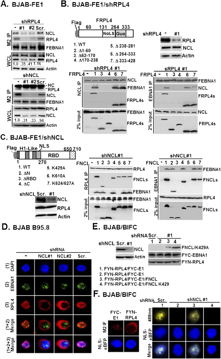
Fig. S5.
RPL4/NCL acts as a protein scaffold for EBNA1. (A) BJAB/FE1/shRL4 #1 or #2 and BJAB/FE1/Scr. control were subjected to M2 IP analysis followed by immune blotting with antibodies for NCL, RPL4, and M2 (Upper). Similarly, BJAB/FE1/shNCL #1 or #2 and BJAB/FE1/Scr. control were used to carry out another set of experiments (Bottom). In both cases, whole-cell lysates were blotted with NCL, RPL4, and internal control actin. (B) The loss of FEBNA1/NCL association in BJAB-FE1/shRPL4 #1 was rescued by transfected FRPL4 or its mutant derivatives followed by co-IP and Western blot analyses. Immune blots for the indicated proteins from NCL and EBNA1-precipitated matrices are shown. Also shown is 2% input of FEBNA and FRPL4 or NCL and FRPL4 from each set of experiments. (C) Same as in B, except BJAB-FE1/shNCL #1- and FNCL-related plasmids were used. Immune blots for the indicated proteins from RPL4 (Middle) and EBNA1 (Right)-precipitated matrices were shown with 2% input of loading control. (D) NCL knockdown by shNCL #1 or shNCL #2 in the context of EBV-positive BJAB B95.8 reversed RPL4 redistribution, whereas Scr. had no effect. EBNA1/RPL4 double immunostaining was carried out, and nuclei were counterstained with DAPI. (E) BJAB cells were cotransfected with FYC-E1 and NLS-eBFP, or FYN-RPL4 and NLS-eBFP. (F) The cellular localization of FYC-E1 and FYN-RPL4 was represented as M2 immunofluorescence images (red), whereas the nuclear regions were marked by NLS-eBFP (blue). BJAB/shNCL #1 cells (columns 2–4) were cotransfected with FYN-RPL4 and FYC-E1, or FNCL and FNCL K429A. The resulting BiFC image in each transfectant was visualized by confocal fluorescent microscopy. The BiFC produced by transfected FYN-RPL4/FYC-E1 in Scr.-transduced BJAB (column 1) was shown.

