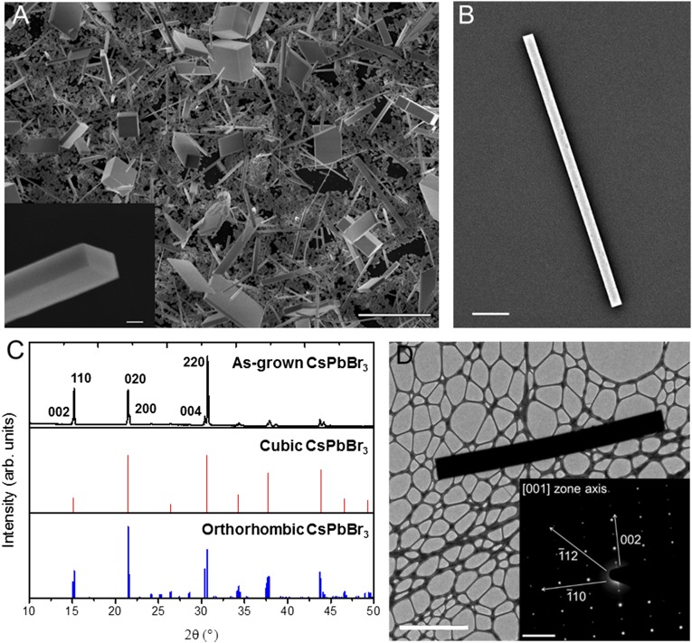
Fig. 1.
Structural characterization of single-crystal CsPbBr3 nanowires. (A) SEM images of CsPbBr3 nanowires and nanoplates grown from PbI2 in a methanolic solution of 8 mg/mL CsBr at 50 °C for 12 h. (Scale bar, 10 μm.) (Inset) SEM image of the rectangular end facet of a nanowire. (Scale bar, 500 nm.) (B) A single CsPbBr3 nanowire isolated on a quartz substrate with a 5-nm Au sputter coat. (Scale bar, 1 μm.) (C) XRD pattern of as-grown CsPbBr3 (black) with the standard XRD patterns of cubic (red) and orthorhombic (blue) CsPbBr3. (D) TEM image of an individual nanowire. (Scale bar, 5 μm.) (Inset) SAED pattern from the same nanowire, with relevant crystallographic axes labeled. (Scale bar, 2 nm−1.)

