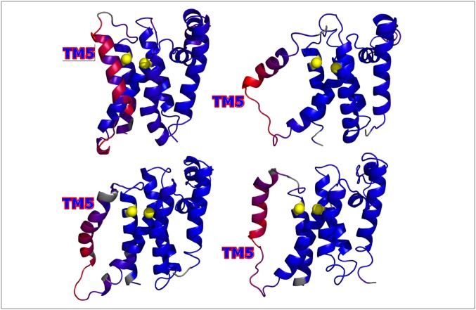
Fig. 6.
Representative structures from a near-native state (F*) (Fig. 5) sampled while simulating with the implicit membrane present. The structures were all aligned to the closed crystal structure (PDB ID code 2XOV) and colored according to the individual residue rmsd values. Blue indicates low rmsd (high similarity to the crystal structure), and red indicates high rmsd. The catalytic dyad is shown using yellow spheres. High rmsd values are localized to the C-terminal half of the molecule and to TM5 in particular. Movement of TM5 exposes the catalytic dyad, thereby allowing substrate access. This state is highly populated under folding conditions when strengthening the contacts in the N-terminal half of the molecule by 10% relative to the contacts in the C-terminal half of the molecule.

