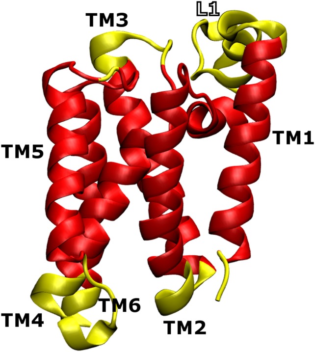
Fig. S1.
Proper topology of the native structure of GlpG used in the implicit membrane model. Residues in the transmembrane region are colored in red. Periplasmic and cytoplasmic residues are colored in yellow. L1 is large and contains two interfacial helices, of which residues 137–143 were assigned to be in the transmembrane region.

