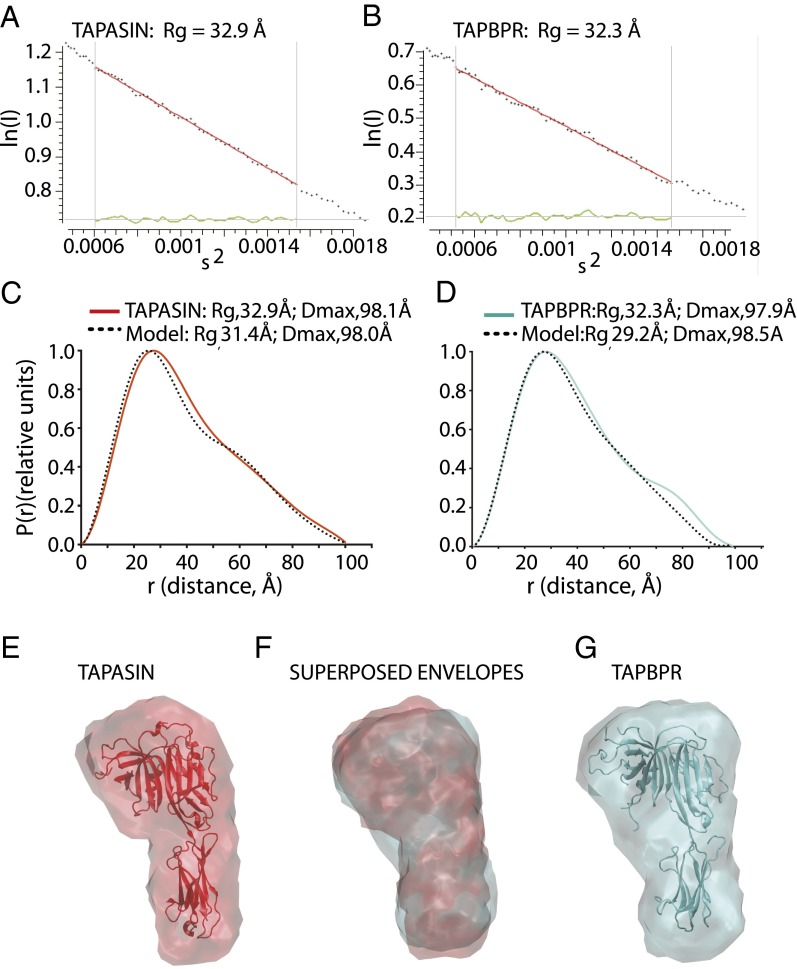
Fig. 6.
SAXS structure determination of tapasin and TAPBPR. Purified recombinant tapasin or TAPBPR were subjected to X-ray scattering data collection upon synchrotron irradiation as described in Materials and Methods. (A) Guinier plot of tapasin scattering data, s Rg limits (0.81–1.29) and (B) Guinier plot of TAPBPR scattering data, s Rg limits (0.75–1.26). (C) Pairwise distance distribution P(r) as a function of r for the experimental data (red) and for a theoretical model for tapasin calculated from the X-ray coordinates (dotted black) (PDB ID code 3f8U). (D) P(r) as a function of r for TAPBPR experimental data (cyan) is compared with a homology model for TAPBPR based on the tapasin structure. (E) Tapasin structure was docked to the SAXS envelope (red), and (G) TAPBPR homology model was docked to its SAXS envelope (cyan). (F) Superposed envelopes of tapasin (red) and TAPBPR (cyan).

