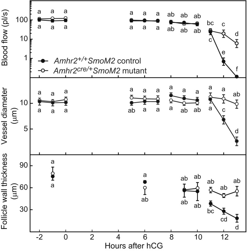
Fig. 2.
Blood flow (Top) and the diameter of vessels within the theca at the apex of the preovulatory follicle (Center) decreased acutely within hours of follicle rupture in Amhr+/+SmoM2 control mice but remained relatively constant in anovulatory Amhr2cre/+SmoM2 mutant mice in which follicular vessels are deficient in VSM. (Bottom) The thickness of the apical follicle wall decreased simultaneously with decreased blood flow in control mice but not in mutant mice. Individual vessels and the thickness of the follicle wall were measured hourly in groups of anesthetized mice studied during 2-h intervals between −2 h to 13 h after injection of hCG. Ovulation occurred in control mice between 13 and 13.5 h after hCG injection. Data on blood flow and vessel diameter at each time point are from 22–36 vessels from individual follicles of five mice (mean ± SEM). Data on the thickness of the follicle wall are from single follicles measured at the times shown in three or four mice. Data points without a common super- or subscript are different (P < 0.05).

