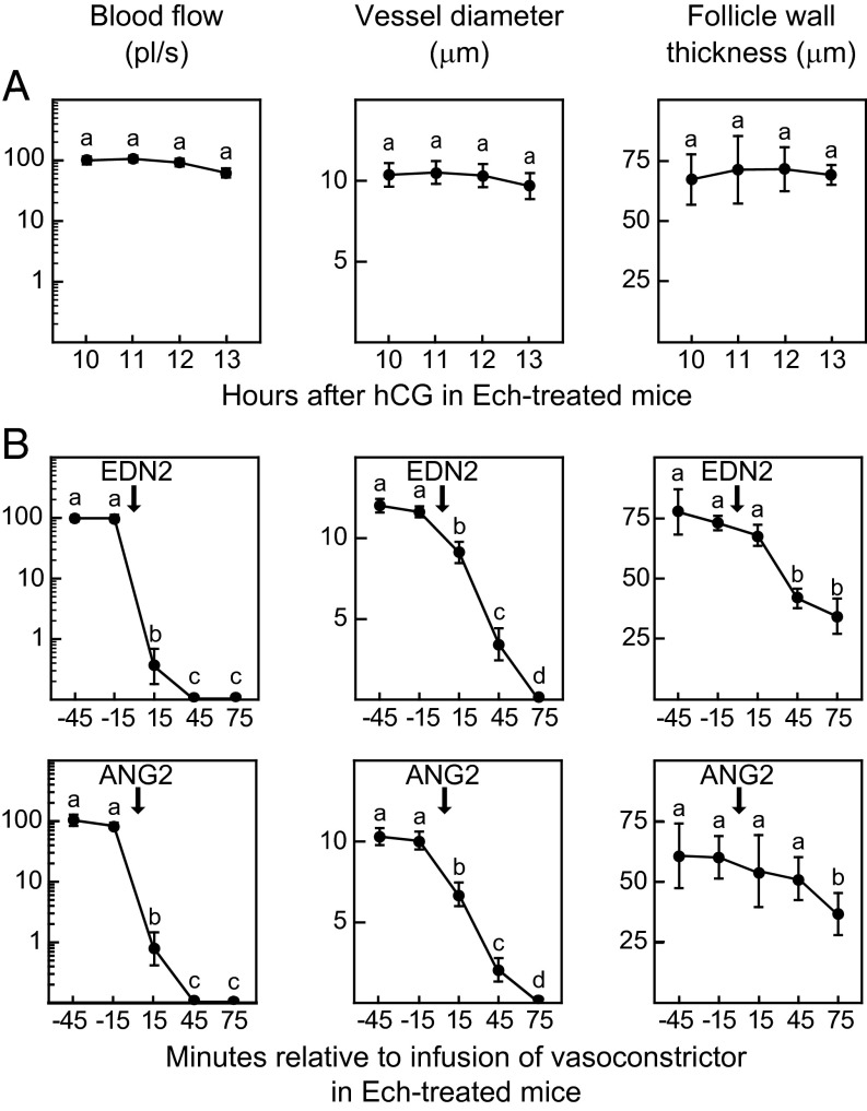
Fig. 4.
Vascular changes within the theca at the apex of preovulatory follicles associated with the suppression of ovulation in Ech-treated wild-type mice and the restoration of ovulation by bursal infusion of vasoconstrictors in Ech-treated mice. (A) Blood flow (Left), the diameter of thecal vessels (Center), and the thickness of the apical follicle wall (Right) were constant from 10 to 13 h after hCG injection in Ech-treated mice in which ovulation was blocked. Data are mean ± SEM from hourly repeated measurements of 17 vessels and of the wall thickness of single follicles from three mice. (B) Infusion of EDN2 (Upper) or ANG2 (Lower) into the bursa of Ech-treated mice restored the changes in thecal vessels and the thinning of the follicle wall that normally occur before rupture. Treatment began at 11.25 h after hCG injection with continuous infusion of 2% BSA-saline for 45 m followed by continuous infusion of vasoconstrictors (arrows) until 15 h after hCG injection. Data are mean ± SEM from 13 EDN2-infused vessels or 15 ANG2-infused vessels and follicle walls imaged repeatedly at 30-min intervals (n = 3 mice for each vasoconstrictor). Data points without a common superscript are different (P < 0.05).

