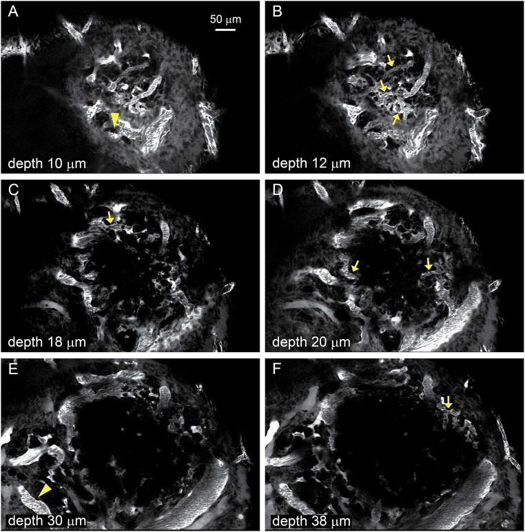
Fig. S1.
A–F show Individual images from a z-stack of the follicle shown as a projection in Fig. 1. The images were obtained at the indicated depths from the surface and show all the thecal vessels (arrows) and vessels external to the theca (arrowheads) analyzed for flow and diameter.

