Fig. S3.
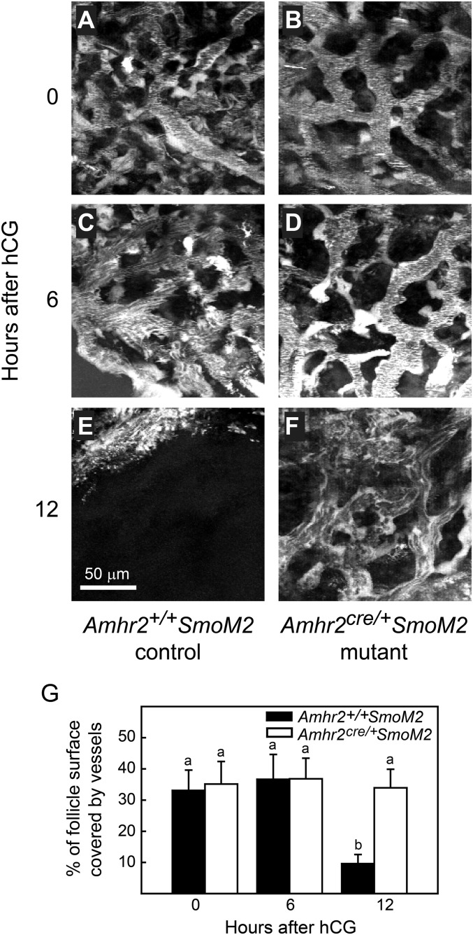
The percent of the surface of preovulatory follicles covered by rhodamine dextran-filled vessels decreased between 6 h and 12 h after hCG injection (just before ovulation) in Amhr+/+SmoM2 control mice but remained constant in anovulatory Amhr2cre/+SmoM2 mutant mice. A single image representing the surface density of vessels was created by projection of 20 sequential images at 2-μm intervals taken from the outer surface of the follicle toward the center. (A–F) Z-stack projections of representative follicles at various time points are shown. A single threshold level was applied to all images to determine the percent of the apical area that was rhodamine dextran-positive and therefore represented filled blood vessels. (G) Data (mean ± SEM) from single follicles of three mice of each genotype are shown. Data points without a common superscript are different (P < 0.05).

