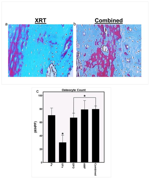
Figure 3.
Histological analysis: (a and b) Representative 16x high power field sections with Gomori’s trichrome stain, demonstrating the diminution of osteocytes in lacunae within irradiated bone, and the comparative sparing of osteocytes observed with combined therapy. (c) ROI osteocyte count metric. *: p < 0.05

