
Figure 14.
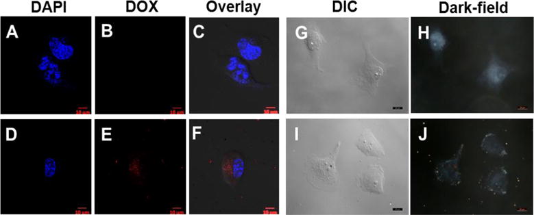
The confocal (A–F) and dark-field (G–J) images of A2058 cells co-cultured with DOX@GoMe. Cells in A–C, and G–H were control. Cells in D–F, and I–J were treated with DOX@GoMe. Images H and J were collected in dark-field mode. Scale bars in A–J are 10 μm.