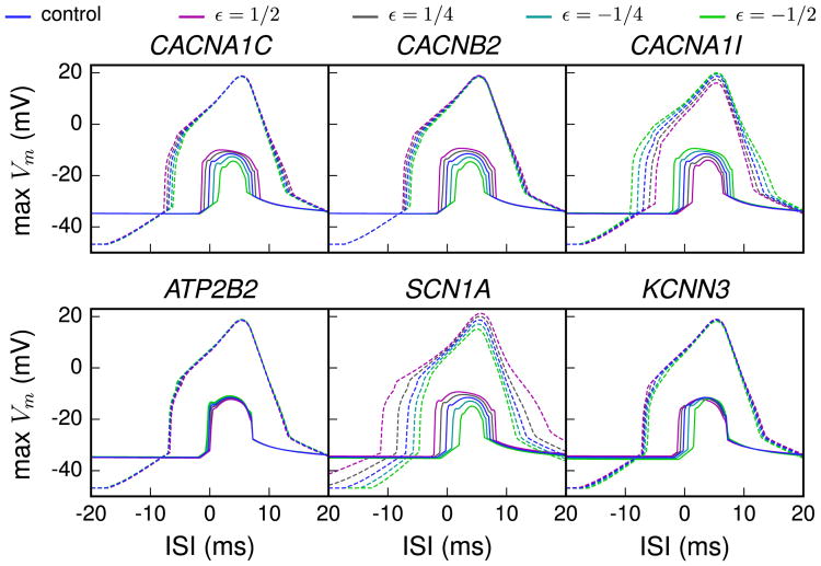
Figure 5.
Sensitivity of variant neurons to interstimulus interval (ISI) between somatic and distal apical stimuli during up and down states. The magnitude of the calcium spikes was assessed by measuring the simulated membrane potential near the bifurcation point of the apical dendrite, i.e., at a distance of 620 μm from the soma. The dashed lines show the temporal maximum of the model response membrane potential during a down state and the solid lines during an up state. The x-axis represents the ISI between the somatic and apical stimuli, positive values denoting cases where the apical stimulus was applied after the somatic stimulus. The variants shown are the same as in Figure 2. In the up state paradigm, the neuron was first given a depolarizing square-pulse current of .42 nA × 200 ms at the proximal apical dendrite, 200 μm from the soma. In the middle of this period, a somatic square-pulse current of .5 nA × 5 ms was applied, and after a time defined as the ISI, an alpha-shaped current injection with rise time .5 ms, decay time 5 ms, and maximal amplitude of .5 nA was applied. The down state paradigm was otherwise equal, but the long depolarizing current was absent, and to compensate for this, the short somatic square-pulse current had amplitude of 1.8 nA.

