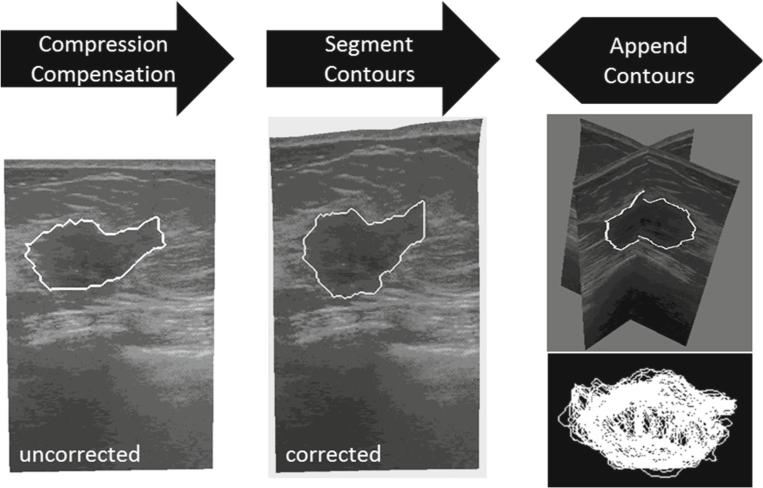
Fig. 6.
Steps involved in processing tracked intraoperative ultrasound data. The ultrasound images are first corrected for tissue compression exerted by the ultrasound transducer. The tumor contour is then seg mented in each 2D slice. Lastly, all contours are appended to form a 3D representation of the intraoperative tumor

