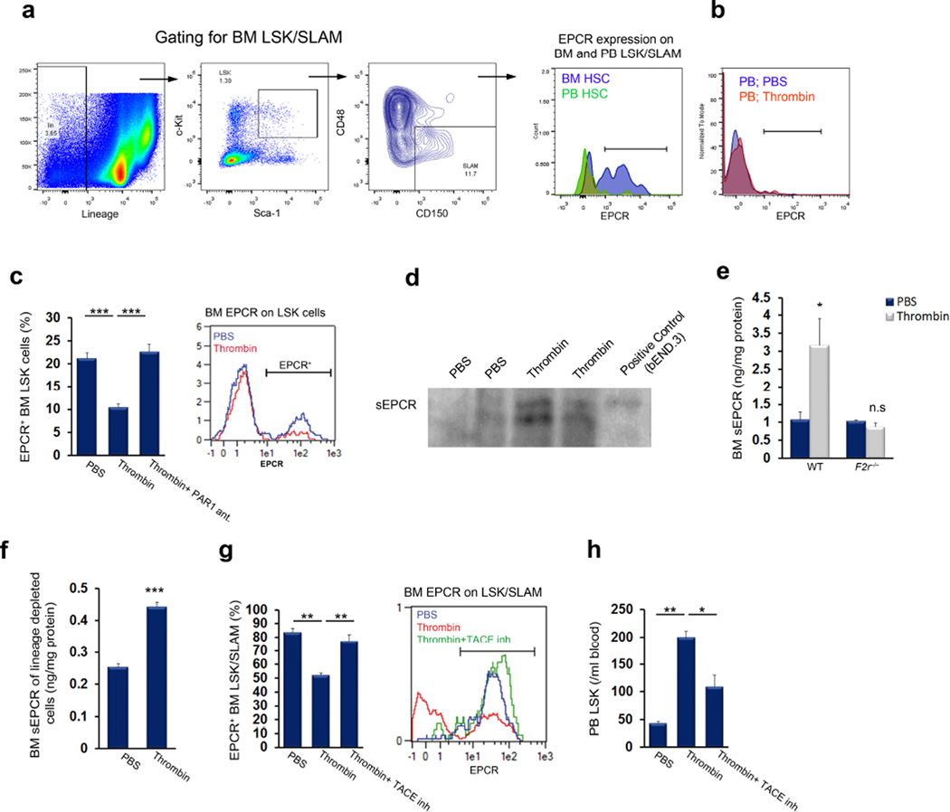
Figure 2. Thrombin-PAR1-dependent EPCR shedding induces HSC mobilization.
(a) FACS analysis of EPCR expression on circulating and bone marrow (BM) LSK SLAM cells; representative data are shown for 4 mice. (b) FACS analysis of EPCR expression on circulating LSK cells in mice treated with thrombin or PBS; representative data are shown for 4 mice. (c) Bone marrow EPCR+ LSK cells following thrombin injection with (n = 4) or without (n = 8) administration of a PAR1 antagonist; P values, one-way ANOVA with Tukey’s post-test. (d,e) Soluble EPCR (sEPCR) in bone marrow fluid 1 hour after thrombin injection, as measured by western blotting (d) or ELISA (e) in wild-type or F2r−/− mice; n = 4. (f) sEPCR ELISA following thrombin stimulation in vitro in lineage-depleted bone marrow cells; n = 4. (g,h) Bone marrow EPCR+ LSK SLAM cells (g) and peripheral blood LSK cells (h) in PBS or thrombin-treated mice, with or without pretreatment with TACE inhibitor for 1 hour before thrombin injection; n = 4; P values, one-way ANOVA with Tukey’s post-test.

