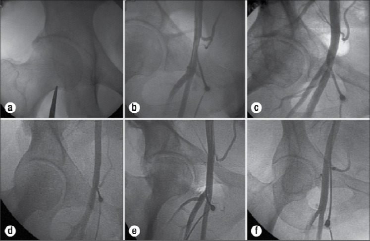
Figure 3.

(a) Fluoroscopy of the femoral head utilizing forceps to note the position of the inferior border of the femoral head on the patient’s skin. (b) Correct placement of the sheath in the common femoral artery. (c) Correct placement of the sheath in relation to the femoral head, with the arterial access incorrectly placed in the superficial femoral artery due to the anatomic variant of a high bifurcation. (d) Correct placement of the sheath in relation to the femoral head with a low hypogastric artery causing incorrect arterial placement in the external iliac artery. (e) Low sheath placement in the profunda femoris artery. (f) High sheath placement in the external iliac artery (Jacobi et al., 2009).
