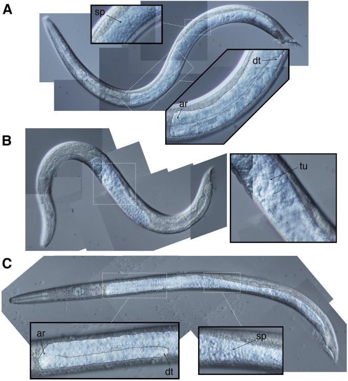
Figure 3.
Gonad morphology in F1 male hybrids. (A) C. nigoni EG5268, (B) F1 XCni, and (C) F1 XCbr males. Contrast of gonads enhanced in all panels. Boxes correspond to regions enlarged in insets. In panels A and C, the distal arm is outlined with a dashed line in the large insets to emphasize the tubular structure of the gonad. This tubular structure is absent in the F1 XCni male shown in panel C. Anterior reflex (ar), distal tip (dt), sperm (sp), and tumorous cells (tu) indicated in insets. The C. F1 XCni male was an ‘exceptional’ GFP– male obtained from crosses on C. nigoni EG5268 males to C. briggsae PB192 [cbr-him-8(v188) I; stIs20120 (pmyo2::GFP) X] hermaphrodites. The F1 XCbr male was a GFP+ male obtained from the same cross.

