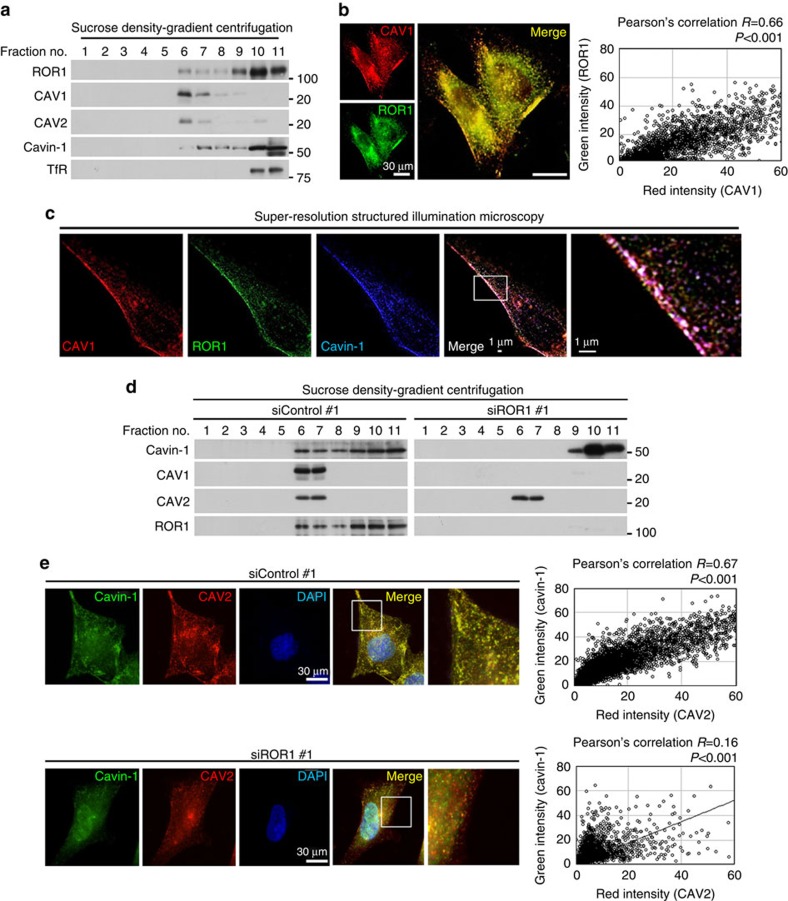
Figure 3. ROR1 colocalizes with CAV1 and cavin-1 and retains cavin-1 in DRM.
(a) Sucrose density-gradient centrifugation confirmed the presence of ROR1 and cavin-1 in the DRM fractions containing CAV1 and CAV2 in the NCI-H1975 cells. (b) ROR1 and CAV1 colocalization shown by two-colour immunofluorescence staining in NCI-H1975 cells. Colocalization was quantified using ImageJ software. Also see Supplementary Fig. 5. (c) The colocalization of ROR1 with CAV1 and cavin-1 shown by three-colour immunofluorescence staining using super-resolution structured illumination microscopy in NCI-H1975 cells. (d) Sucrose density-gradient centrifugation showing the loss of CAV1 as well as marked changes of cavin-1 subcellular distribution in NCI-H1975 cells knocked down for ROR1. Also see Supplementary Fig. 6a. (e) Two-colour immunofluorescence staining showing markedly impaired colocalization between cavin-1 and CAV2 induced by ROR1 knockdown in NCI-H1975 cells. Colocalization was quantified using ImageJ software. Also see Supplementary Fig. 6b. Uncropped images of blots are shown in Supplementary Fig. 11.

