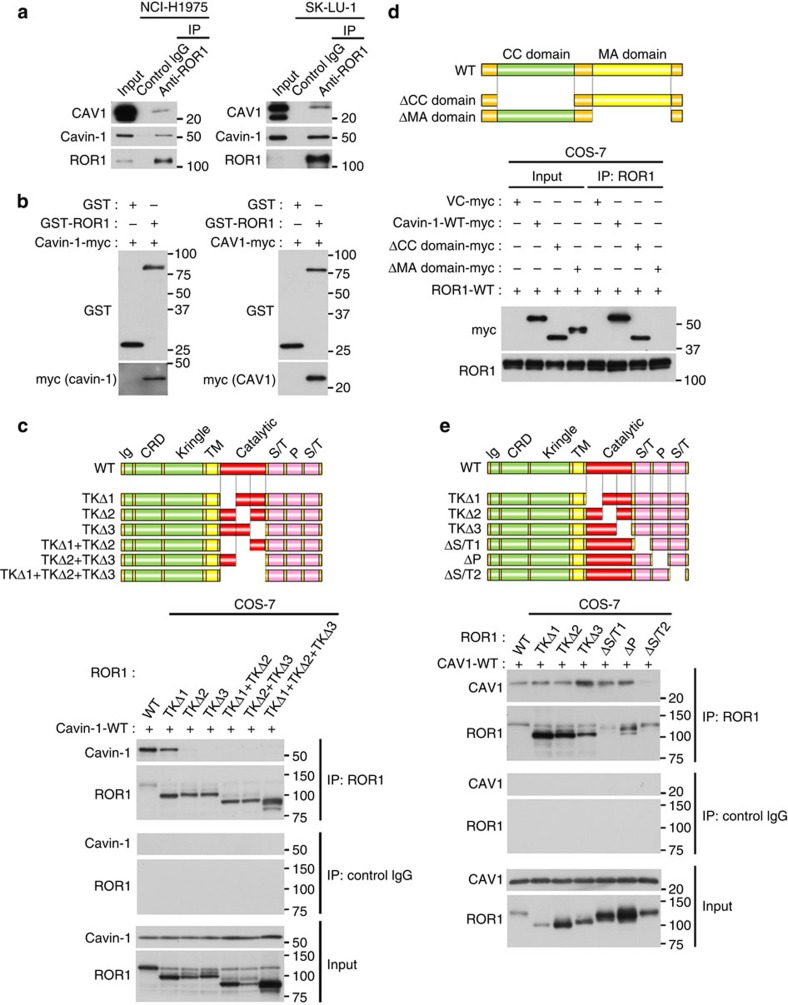
Figure 5. ROR1 interacts with both cavin-1 and CAV1 at two respective binding regions.
(a) IP–WB analysis using octylglucoside as a detergent showed association of ROR1 with cavin-1 and CAV1. See Supplementary Fig. 8a. Also see Supplementary Fig. 8b for pull-down assay showing their mutual associations. (b) Pull-down assay using purified proteins of ROR1, cavin-1, and CAV1 showing physical associations of ROR1 with cavin-1 and CAV1. (c) Identification of C-terminal two-thirds of the ROR1 kinase domain as the cavin-1 binding region by IP-WB analysis using various ROR1 deletion mutants. Also see Supplementary Fig. 8c–f. (d) Identification of the membrane association domain of cavin-1 as its ROR1-binding region by IP–WB analysis using cavin-1 deletion mutants. Also see Supplementary Fig. 8g. (e) Mapping of the CAV1 binding region to the C-terminal serine/threonine-rich domain by IP–WB analysis using various ROR1 deletion mutants. Uncropped images of blots are shown in Supplementary Fig. 11.

