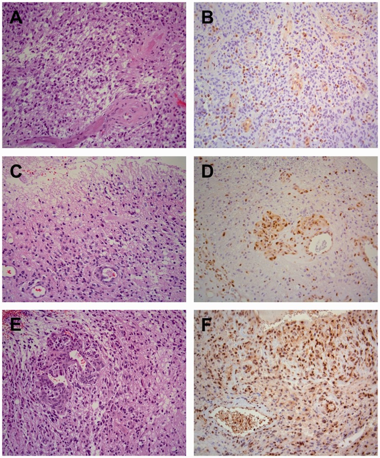
FIGURE 3.
Representative examples of MGMT immunohistochemistry (IHC) on glioblastomas (World Health Organization [WHO], grade IV). (A, C, E) There are characteristic pleomorphic glial tumor cells, necrosis with pseudopalisading and endothelial cell proliferation. Hematoxylin and eosin stain. (B, C, F) Variable amounts of staining by IHC for MGMT of endothelial cells, lymphocytes, microglia, and tumor cells. (B) Only a minority of tumor cells is positive (0%–10%). (D) There is clear staining of endothelial cells in tumor vessels and diffusely in tumor cells (11%–25%). (F) Many endothelial cells and tumor cells are positive (26%–50%). The staining of tumor cells is heterogeneous, varying from area to area in the tissue. Magnification: x200 for all panels.

