
Figure 1.
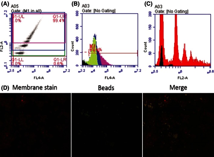
(A) FL4‐A represents CD14+ events, FL2‐A represents PE + events. Distinct populations of cells engulfing 1, 2, and 3+ beads can be seen ascending in PEfluorescence. (B) CD14+ events are separated to show cells that are engulfing 1 bead (green), 2 beads (blue), and 3+ beads (pink). (C) PE + events overlayed with CD14+ in red. The 1 bead population shows slight contamination with nonCD14+ events (black). This may be residual B cells which might have beads attached on the membrane. The 1 bead population was not used in analysis because of this contamination. (D) Mononuclear phagocytes postaddition of beads. We can observe there are very few beads on the membrane or free beads after 1 h incubation and washes. Most beads are fully internalized.