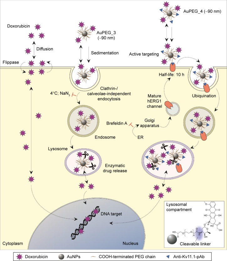
Figure 13.
The proposed mechanism of cell internalization of PEG-AuNPs (AuPEG_3 and AuPEG_4) and drug release compared to molecular DOX are shown.
Notes: PEG-AuNPs reach the cell membrane by diffusion and, in vitro, by sedimentation. Following association to cell membrane, AuPEG_3 is internalized by clathrin-/caveolae-independent endocytosis. Endosomes are then merged with lysosomes, where DOX is released by the enzymatic cleavage of the amide bond through which DOX molecules are grafted to the PEG layer. In parallel, AuPEG_4 associates to the cell surface by active targeting of the Kv11.1 subunit of the hERG1 channels. Mature hERG1 K+ channels remain at the plasma membrane with a half-life of ~10 hours.99,100 During degradation, hERG1 channels are internalized in endocytic vesicles, tagged with ubiquitin, and then degraded in lysosomes.101 Here, DOX is enzymatically released from AuPEG_4. Once released within the cell cytoplasm by diffusion, DOX can reach the nucleus and bind to target DNA. Internalization of molecular DOX relies on diffusion of the drug to the target cell, the association/dissociation equilibrium to/from the cell membrane, and the transport from the inner to outer leaflet (and back) by flippases. AuNPs are represented by gray circles, PEG chains by the (~) symbol, DOX by purple stars, and the anti-Kv11.1-pAb by blue triangles. “T”-shaped symbols indicate inhibitors. Please note that drawings are not to scale.
Abbreviations: PEG, polyethylene glycol; AuNPs, gold nanoparticles; DOX, doxorubicin; AuPEG, PEG-coated AuNPs; h, hours.

