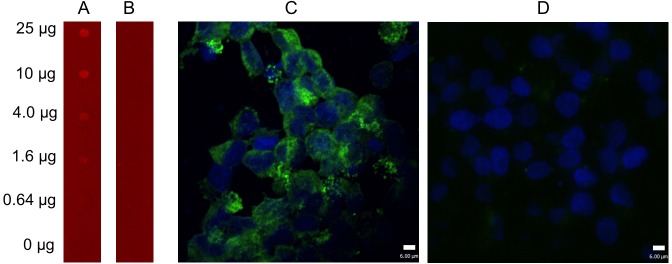
Fig 4. Analysis of binding of epitope 1 to MAb D7 against NS1.
The results of A dot-blot immunoassay assay (A and B) and confocal laser scattering microscopy images (C and D). The peptide GSGSDAPFGSGS was spotted on the activated membranes and incubated with anti-NS1 MAb D7 (A) and control serum (B), staining of infrared-labeled goat anti-mouse IRDye 700 secondary antibody. The results showed that the peptide reacted to the MAb D7 in a concentration-independent manner. In addition, 293T cells were transfected with pCAGGS-NS1 (C) and pCAGGS-NS1-del-DAPF (D) using Lipofectamine 2000. After 36 h, 293T cells were fixed and probed with mouse anit-NS1 MAb ascitic fluid (dilution 1:100) followed by the FITC-conjugated goat anti-mouse antibody (green). Nuclei were counterstained with DAPI (blue). Scale bars are 6 μm.

