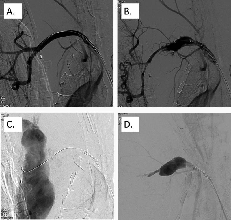
Fig. 5.
Baseline angiographic scan of the right A. axillaris (A). Dis-ruption of the right A. axillaris using a balloon dilation (B). Further disruption of the right A. axillaris using balloon dilation to increase blood loss dynamics (C). Angiographic scan 17 min after the admin-istration of rFVIIa through the drawn back angiography catheter,demonstrating a contained hematoma without active extravasation of blood (D).

