
Figure 3.
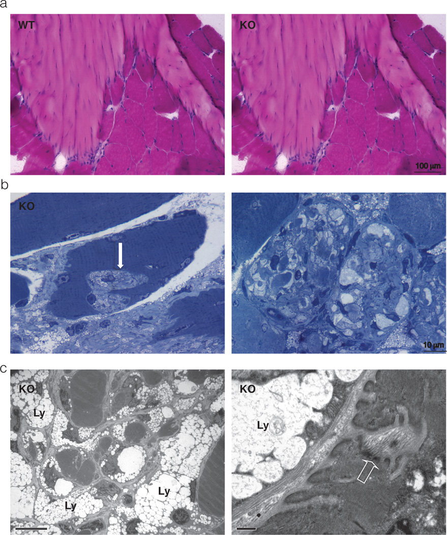
a) Hematoxilin & Eosin staining of transverse sections of WT and Neu1−/− gastrocnemius muscle; large areas of connective tissue infiltrates and increased number of cells in the Neu1−/− myotendinous junction are observed when compared to the WT tissue; b) Toluidine blue-stained semithin Neu1−/− gastrocnemius muscle sections; invagination of the sarcolemma with infiltration of the muscle fibers by ECM components is observed (arrow on the left panel); in an advanced stage, ECM invagination results in severe fragmentation of the cytosol and complete disruption of the muscle cytoarchitecture as observed in a transverse section (right panel); c) Electron microscopy ultrastructural analysis of Neu1−/− gastrocnemius muscle, showing cytosolic fragmentation and infiltration of muscle fibers by fibroblast-like cells whose cytosol is filled with storage lysosomes (Ly) and thickening of the sarcolemma (open arrow). Adapted from Zanoteli et al. Biochim, Biophys Acta 2010, with permission of Elsevier.