
Figure 3.
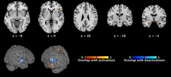
Topographical overlap between present functional connectivity maps of areas with pedophilia‐related neuroanatomical alterations and previously meta‐analytically determined regions implicated in sexual arousal. Conjunction maps of significant overlap between the present (FWE corrected) functional connectivity maps of each seed (cf. Fig. 1) and the previously published (FWE corrected) ALE maps of sexual arousal [Poeppl et al., 2014]. Sagittal, coronar, and axial brain slices are shown at MNI coordinates (x, y, z). ALE, activation likelihood estimation; MNI, Montreal Neurological Institute. [Color figure can be viewed in the online issue, which is available at http://wileyonlinelibrary.com.]