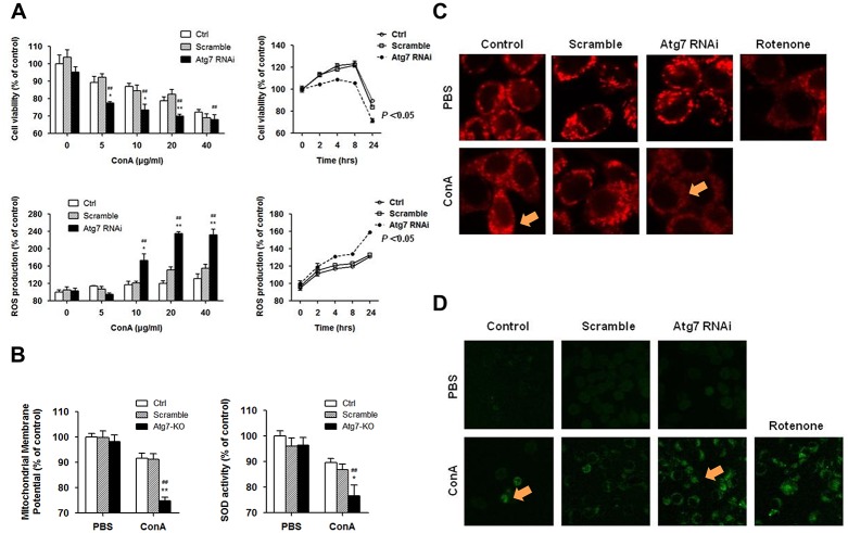
Fig 3. Atg7 silencing resulted in accumulation of dysfunctional mitochondria and abolished ROS degradation in Raw264.7 cells.
A. Cell viability measured by an MTT assay and ROS production by mean fluorescence intensity of H2DCF-DA at different concentrations and time points for ConA exposure in Raw264.7 cells lacking Atg7. B. Measurement of mitochondria membrane potential and SOD activity after ConA stimulation. Raw264.7 cells were transfected with Atg7 siRNA or control siRNA and treated with 10 μg/ml ConA for 24 h. Representative results were from 3 independent experiments. Data are presented as mean±SD; * p<0.05 and ** p<0.01 vs. controls, # p<0.05 and ## p<0.01 vs. pre-treatments. C and D. Fluorescence microscopy of mitochondrial membrane by Mito-tracker Red FM (red) and ROS production by H2DCF-DA (green) after ConA stimulation (Arrows showing fluorescence staining regions). Raw264.7 cells were transfected with Atg7 siRNA or control siRNA and treated with 10 μg/ml ConA for 4 h. Magnifications: 1000X. Representative results were from 3 independent experiments.

