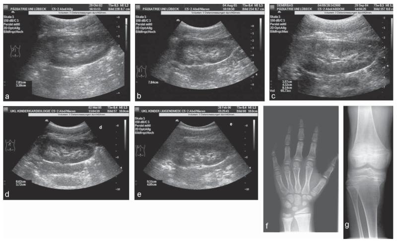
Fig. 2.
Ultrasound of the left kidney and X-rays of individual II-4 homozygous for the G196R mutation. Panels a to e show the renal ultrasound (a: age 8; 10 years, b: age 9; 8 years, c: age 10; 9 years, d: age 11; 5 years, e: age 12; 2 years). Panel a was taken before initiation of therapy and demonstrates slight nephrocalcinosis corresponding to grade I, nephrocalcinosis worsens slightly in due course to grade IIa (panels b to e). Panel f demonstrates the X-ray of the hand before therapy (age 8; 5 years) with a bone age of 6; 6 years (Greulich and Pyle) and shows slight flaring and mild osteopenia apart from brachymesophalangia. Panel g shows the X-ray of the right knee (age 11; 5 years) with epiphyseal flaring, numerous transverse striations in the metaphysis and Erlenmeyer flask deformities in the metaphysic.

