Figure 1.
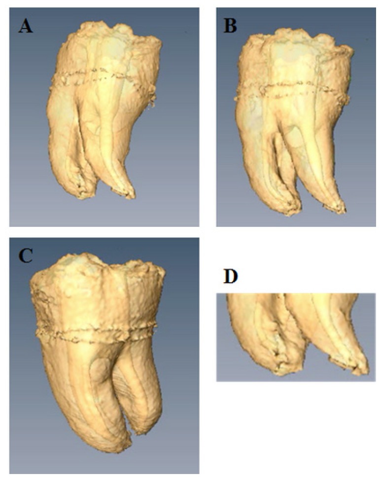
3D volume reconstruction of the internal and the external morphology of an endodontically treated third molar.
A. Distal-vestibular view of the distal root and root canal shaped at a 6% taper. An edifying image of the prepared root canal provided by MRI. A clear view upon the irregular shape of the prepared distal root canal, meaning that a part of the complex anatomy of the distal root canal was left untouched, i.e., not cleaned by the mechanical shaping files.
B. Vestibular view of the external morphology of mesio vestibular and distal roots and the correspondingly shaped root canals. Root canal curvatures, the furcal region, the interradicular root grooves can be clearly seen.
C. Proximal (mesial) view. Note the degree of root separation and the apical fusion of the two mesial roots.
D. Detail of the apical part of the three roots and root canals. The root canal openings (portal of exits), the apical finishing of the root canal treatment can be viewed with great accuracy.

