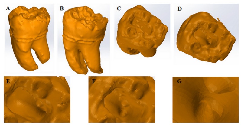
Figure 2.
3D volume rendered filled surface reconstruction.
A. External Lingual view;
B. External Vestibular view;
C. Occlusal view of the pulp chamber;
D. Access Cavity;
E. View of the distal canal opening, on the pulp chamber floor;
F. Distal Root canal opening (close-up);
G. View inside the coronal third of the root canal.

