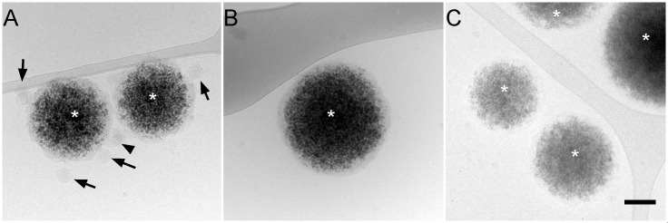
Fig 3. Immunocapture of BVDV particles.
Protein G-coated magnetic beads (asterisks) were incubated with anti-BVDV E2 mAbs (A), no antibody (B), or an irrelevant mAb (C) and purified BVDV fraction and observed by cryo-electron microscopy. Black arrows: enveloped, capsid-containing particles of ~50 nm, black arrowheads: larger enveloped, capsid-containing particles. Bar, 100 nm.

