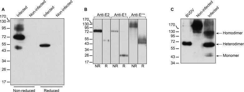
Fig 4. Envelope glycoprotein dimerization.
(A) Triton-X100 lysates of infected and non-infected MDBK cells analyzed by immunoblot with a mix of anti-E2 mAbs WB214 and WB166 under reduced or non-reduced conditions. (B) Purified BVDV analyzed by immunoblot with anti-E2 mAbs WB214 and WB166, anti-E1 mAb 8F2 or anti-Erns pAb under reduced (R) or non-reduced (NR) conditions. (C) Lysates of purified BVDV and infected MDBK cells containing similar amounts of E1E2 heterodimer, and a lysate of non-infected MDBK cells were analyzed by immunoblot with anti-E2 mAbs under non-reduced condition. The bands of E2 monomer, E2 homodimer and E1E2 heterodimer are indicated.

