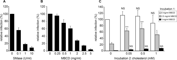
Fig 9. Sphingomyelin and cholesterol in BVDV entry.
(A) Unpurified BVDV was incubated for 1h at 37°C in the presence of indicated concentrations of sphingomyelinase (SMase), diluted 1,000 times and used to infect MDBK cells. (B) Partially purified BVDV was incubated at 37°C for 1 h with indicated concentrations of methyl-β-cyclodextrin (MBCD), diluted 10,000 times and used to infect MDBK cells. (C) Partially purified BVDV was incubated at 37°C for 1 h with indicated concentrations of MBCD (incubation 1), diluted 100 times, incubated for 1h at 37°C with MBCD:cholesterol complex at final cholesterol concentrations indicated (incubation 2), diluted 100 times and used to infect MDBK cells. The number of infected cells was measured using an immunofluorescence assay at 15 hpi and standardized to the number of cells infected with untreated virus. Error bars correspond to SDs (n = 3, * P<0.05, *** P<0.001, NS: P>0.05; cholesterol-treated vs corresponding control, 2-way ANOVA).

