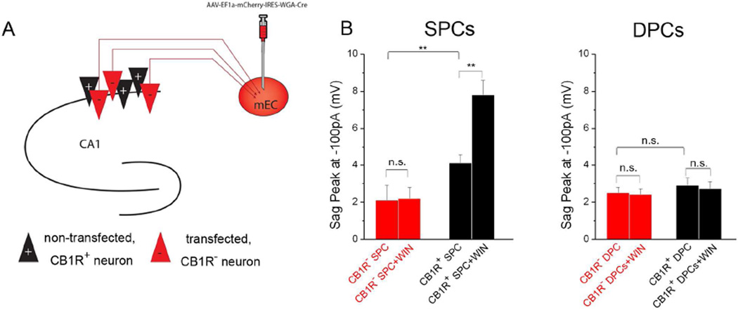
Figure 4. Postsynaptic CB1Rs modulate Ih in SPCs.
A) Experimental paradigm. mEC: medial entorhinal cortex; Red triangle: cells expressing the viral vector, lacking CB1R (CB1R− cells); Black: uninfected, CB1R+ cells.
B) Peak sag amplitudes recorded at −100 pA in CB1R− and CB1R+ SPCs (left) and DPCs (right) before and after WIN application (n: CB1R− SPCs: 10; CB1R− SPCs + WIN: 15; CB1R+ SPCs: 13; CB1R+ SPCs+WIN: 15; CB1R− DPCs: 11; CB1R− DPCs+WIN: 13; CB1R+ DPCs: 14; CB1R+ DPCs+WIN: 13). The effect on sag amplitude was similar for all current injections between −400 and −100 pA (Tables S1 and S2).

