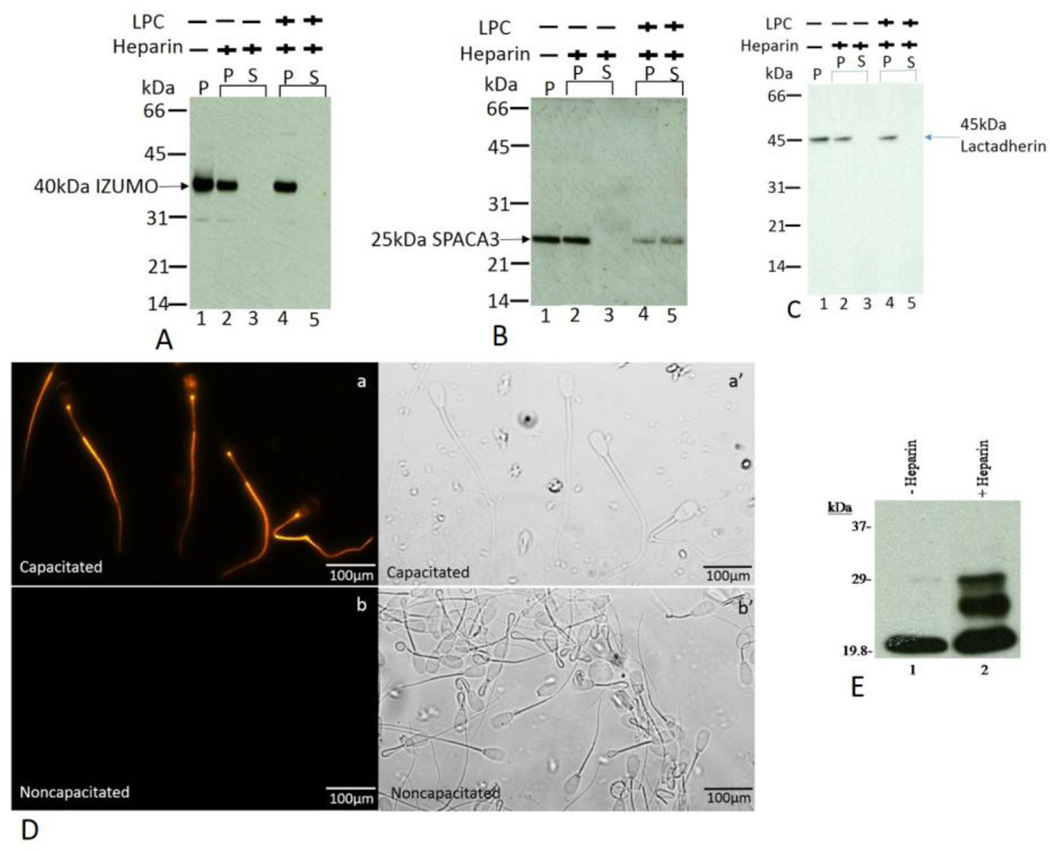
Figure 7.
A, B, and C: Immunoblot analysis of the pattern of IZUMO1 (Fig. A), SPACA3 (Fig. B), and lactadherin (Fig. C) polypeptides in the particulate (P) and supernatant (S) fractions of spermatozoa after a 4 hr incubation in the presence or absence of heparin and with or without a final 15 min exposure to LPC. The immunoblot was stained with anti-IZUMO1 (Fig. A), anti-SPACA3 (Fig. B), anti-lactadherin (Fig. C). Note that under each treatment condition the entire IZUMO1 and lactadherin pools remain in the pellets (Figs. 7A and 7C, lanes 1, 2 and 4); no immunoreactive band was detected in the supernatant fractions (Figs.7A and 7C, lanes 3 and 5). On the contrary, as demonstrated in Figure 7B (lanes 1 and 2), the entire SPACA3 polypeptide was retained in the pellet of noncapacitated and capacitated sperm pellet fractions and a significant portion of SPACA3 was released after LPC-induced acrosome reaction (Fig.7B, lane 5). The remaining portion of SPACA3 was retained in the sperm pellet (Fig.7B, lane 4).
D: Immunocytochemical localization of tyrosine-phosphorylated proteins in capacitated (panel a and a’) and noncapacitated spermatozoa (panel b and b’). Matched phase contrast (panels a’ and b’) and fluorescence (panels a and b) photomicrographs of spermatozoa stained with (anti-PY20).
E: Protein tyrosine-phosphorylation pattern of cauda epididymal spermatozoa. Western blots of noncapacitated (lane1) and capacitated (lane 2) spermatozoa fractionated by reducing SDS-PAGE and stained with anti-phosphotyrosine (anti-PY20).

