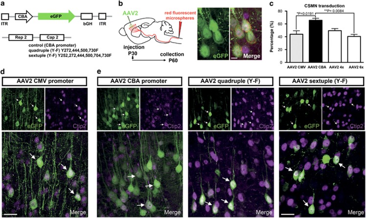
Figure 3.
AAV2-2 containing CBA promoter has high tropism for CSMN. (a) Schematic representation of an AAV cis-shuttle plasmid expressing eGFP under the CBA promoter and trans-plasmid encoding for AAV2-2 rep proteins and capsid. Modified capsids used in this study contained four (AAV quadruple Y-F) and six (AAV sextuple Y-F) tyrosine to phenylalanine point mutations. (b) Experimental approach showing direct injection of AAV2-2 and simultaneous injection of red fluorescent microspheres into the CST. All injections were performed at P30, and the tissue was collected at P60. Representative image of AAV2-2-transduced neurons that are also retrogradely labeled with red fluorescent microspheres (orange-red punctate) and immunostained for Ctip2 (purple) confirming their CSMN identity. (c) Bar graphs represent mean percentage ±s.e.m. of CSMN (eGFP+ cells, expressing Ctip2) transduction with different AAV2-2. (d) Representative images of neurons transduced with AAV2-2 encoding eGFP under the CMV promoter. Arrows indicate eGFP+ neurons expressing Ctip2. (e) Representative images of neurons transduced with AAV2-2 encoding eGFP under the CBA promoter (left), AAV2-2 encoding eGFP under the CBA promoter containing four (AAV2-2 quadruple Y-F) tyrosine to phenylalanine point mutations (middle panel) and AAV2-2 encoding eGFP under the CBA promoter containing six (AAV2-2 sextuple Y-F) tyrosine to phenylalanine point mutations (right panel). Arrows indicate eGFP+ neurons expressing Ctip2. One-way ANOVA and Tukey's multiple comparison test. *P<0.05, **P<0.01. Scale bar=20 μm.

