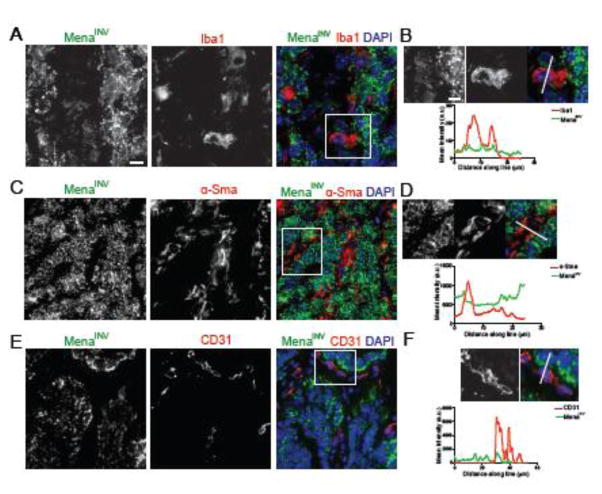
Figure 4. MenaINV expression is restricted to the tumor compartment.
Representative images of staining for MenaINV and the macrophage marker Iba1 (A,B), the fibroblast marker αSMA (C,D) and the endothelial cell marker CD31 (E,F). B,C,D show magnification of boxed area and signal intensity for both markers is quantified along the white line. Scale bar for A,C,E is 30 μm and 10 μm for B,D,F.

