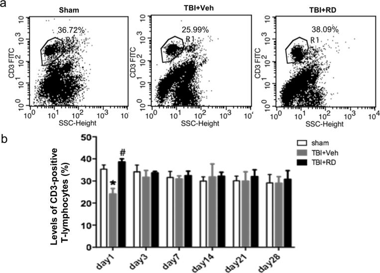
Fig. 4.
Effects of Rhizoma drynariae (RD) on CD3-positive T lymphocytes in the blood of rats subjected to CCI. CD3-positive T lymphocytes in blood were measured by flow cytometry on days 1, 3, 7, 14, 21, and 28 after CCI. a Representative flow cytometric dot plots show the percentage of CD3-positive T lymphocytes in sham, vehicle-treated, and R. drynariae-treated groups on day 1 after CCI. b Histograms show that the percentage of CD3-positive T lymphocytes was lower in the vehicle-treated group than in the sham group on day 1 after CCI and that R. drynariae treatment reversed this decrease. On days 3, 7, 14, 21, and 28 after CCI, the percentage of CD3 T lymphocytes had returned to normal, and R. drynariae treatment had no further effect. Values are mean±SD; n=6 rats/group; *p<0.05 vs. sham group, #p<0.05 vs. vehicle group

