Figure-3.

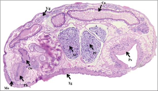
Microscopic picture of medial section of Paramphistomum cervi showing different organs: Mouth (Mo), pharynx (Ph), tegument (Tg), caecum (Ca), vitelline gland (Vg), anterior testis (At), posterior testis (Pt), posterior sucker (Ps).

Microscopic picture of medial section of Paramphistomum cervi showing different organs: Mouth (Mo), pharynx (Ph), tegument (Tg), caecum (Ca), vitelline gland (Vg), anterior testis (At), posterior testis (Pt), posterior sucker (Ps).