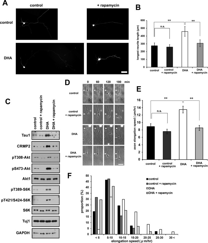
FIGURE 4.
Rapamycin abrogates DHA-induced axon outgrowth. A, EGFP-transfected neurons were treated with or without 1 μm DHA in the presence of rapamycin and fixed at 3 DIV. Representative images are shown. Scale bar = 50 μm. B, longest neurite (axon) measurement (n = 4; control, 55 neurons; control +rapamycin, 72 neurons; DHA, 53 neurons; DHA + rapamycin, 48 neurons). Error bars indicate mean ± S.E., one-way ANOVA with Tukey-Kramer post hoc test. *, p < 0.05; **, p < 0.01; n.s., not significant. C, Western blotting analysis of cell lysates prepared from neurons treated with or without DHA in the presence of rapamycin for 3 DIV. D, rapamycin abrogated the enhanced elongation rate of axons by DHA. Representative time-lapse images are shown. The neurons were imaged at 0, 60, 120, and 180 min. The positions of the growth cone of the axon at 0 min (arrowheads) and at 180 min (arrows) are marked. Scale bar = 25 μm. E, quantification of axon elongation rates on the basis of the time-lapse images shown in D. Error bars indicate mean ± S.E. One-way ANOVA with Tukey-Kramer post hoc test. *, p < 0.05; **, p < 0.01. F, distribution of axon elongation speed. The sum of five independent experiments is shown (n = control, 41 neurons; control + rapamycin, 38 neurons; DHA, 47 neurons; DHA + rapamycin, 44 neurons).

