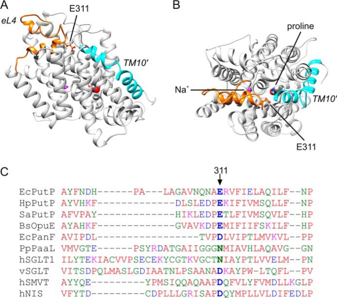
FIGURE 1.

Structural model of PutP. A and B, homology and restraint-based model of PutP in an inward-open conformation with eL4 highlighted in orange and TM10′ in turquoise (A, top view; B, side view). The model represents an advancement of earlier models (28, 29), and matches nine DEER-based distances (root mean square deviations for all distance restraints = 0.72 Å). The side chain of Glu-311 is shown in ball-and-stick representation. The indicated putative locations of the binding sites for l-proline and sodium were taken from Olkhova et al. (28). C, sequence alignment of amino acids of eL4 of SSS family members. The alignment was performed with Clustal Omega (53).
