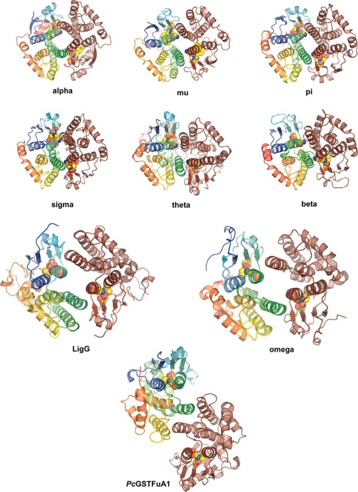
FIGURE 4.
Representative cytosolic GST dimer forms. Representatives from several GST classes are shown, in which one molecule of the dimer is shown in brown, the second molecule of the dimer is shown in rainbow colors (N terminus in blue to C terminus in red), and the bound glutathione or glutathione analog is shown as yellow spheres. The Alpha (Protein Data Bank entry 1GUH; human GST A1-1), Mu (2GST; rat), Pi (2GSR; pGST P1-1 from pig), Sigma (1GSQ; squid), Theta (1LJR; human hGST T2-2), Beta (2PMT; bacterial GST from P. mirabilis), Omega (3LFL; human GST Omega-1), and LigG (4G10; Sphingobium sp. SYK-6) dimers show variations on the α3/α4 canonical four-helix bundle dimer structure, whereas the GSTFuA structure from P. chrysosporium shows a non-canonical dimer formed via interaction between α4 and the C-terminal domain of the second molecule of the dimer.

