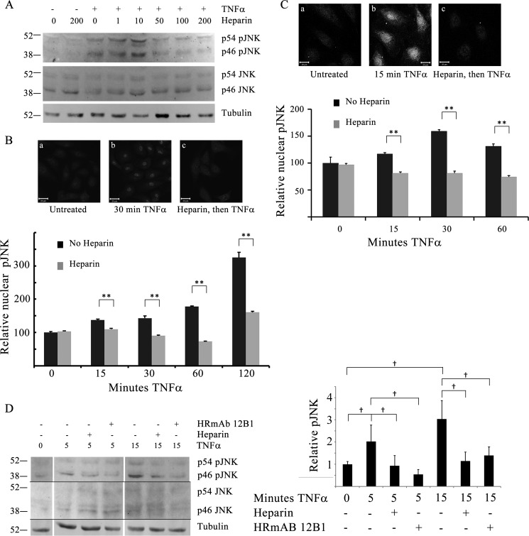
FIGURE 2.
Active JNK is decreased in heparin-treated endothelial cells. BAOECs were treated with 25 ng/ml TNFα for various times. Identical samples were pretreated with 1–200 μg/ml heparin 20 min before TNFα addition. A, cells treated with varying heparin concentrations were harvested into sample buffer and analyzed by Western blotting. Data are representative of more than three repeats. B, to evaluate pJNK in the nuclei, BAOECs were cultured on coverslips and treated as cells in A, and processed for immunofluorescence as described under “Experimental Procedures.” Representative images for one time point are shown. Mean nuclear staining of more than 200 cells/point (from three separate experiments) is shown, with error bars as mean ± S.E. **, p < 0.001. C, HBMECs were treated and analyzed, as were BAOECs. Differences at all times after TNFα addition were significant. **, p < 0.001. D, BAOECs were treated as in A, but some cells were treated with HRmAb 12B1 at 1.0 μg/ml or 200 μg/ml heparin. The space between time points in the image is due to removal of a marker lane and different loading patterns. The graph represents an analysis from four different experiments for the p46 JNK band only. †, p < 0.05.

