
Fig. 2.
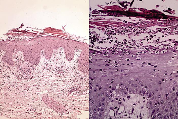
a Epidermis with acanthosis, parakeratosis and moderate perivascular inflammatory infiltrate in the superficial dermis. b Higher magnification showing epidermis with exocytosis of neutrophils and small pustule formation in the stratum corneum.