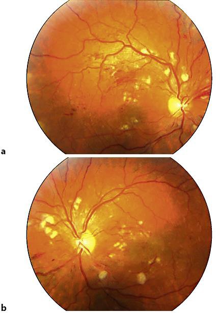
Fig. 1.

a, b Colour pictures of both eyes: ‘flame-shaped’ haemorrhages and hard exudates in the posterior pole, ‘cotton wool spots’ located around the optic disc and disseminated foci of hyperpigmentation arranged linearly on the periphery, indistinct optic disc borders.
