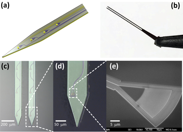
Figure 1.
(a) Schematic of a hypothetical neural probe with an array of optical waveguides, grating couplers and microelectrodes for high spatiotemporal optogenetic stimulation and electrophysiological recording. (b) Photo of a completed double-shank probe held by a tweezer. (c) Microscope image of output grating couplers and a waveguide on a double-shank probe. (d) Microscope image of a probe tip with a grating coupler connected to a waveguide. (e) SEM image of a grating coupler with a length of 15 μm and width of 20 μm.

