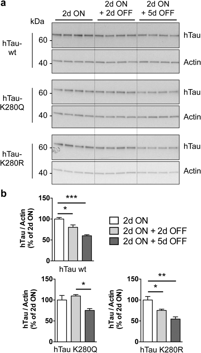
Figure 6. hTau clearance was affected by K280 pseudo-acetylation.
Western blot analysis (a) and quantification (b) of total hTau levels retrieved from adult fly head extracts following neuronal (elavGS-driven) expression of hTau-wt, hTau-K280Q or hTau-K280R following either 2 days of RU486 induction (“2d ON”) or 2 days of RU486 induction followed by exposure to RU486-free food for either 2 or 5 days (“2d ON + 2d OFF” and “2d ON + 5d OFF”, respectively). hTau detection was achieved using the polyclonal K9JA antibody (Dako) and Actin was used for normalisation. Results are expressed relative to levels observed in the “2d ON” condition (*p < 0.05, **p < 0.01 and ***p < 0.001, one-way ANOVA followed by Tukey’s post hoc test, n = 4/condition).

